 Most dental cleanings take between 30 minutes and an hour. If the cleaning is part of a yearly check-up, the dentist may also take x rays by dental x ray machine to check for cavities. A dental hygienist usually performs most of the cleaning. The dentist will normally take a last look, and perhaps perform some difficult plaque removal toward the end of the cleaning, as well as evaluate the gums for gum disease.
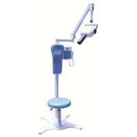
There are specialised machines available for cleaning and lubrication of drills. Yep, the good old dental drill is pricey enough to warrant its own cleaning system, and each dental practice requires a bunch of drills so they can be clean for each patient. Next time you visit the dentist you may also notice the amount of disposable items that are used to stop infection spreading from patient to patient. Dental hygienists generally use several tools during a dental cleaning, including a tooth polisher and a scaler. Tooth polishers buff teeth and eliminate tiny pieces of plaque. When a dental hygienist cleans and polishes your teeth, you can get a lot of cleaning paste in your mouth. We use the suction to help clean all of that away. Also, when dentists are do amalgam fillings( dental amalgamator ), pieces of the soft amalgam can sometimes fall away from the tooth surface. If you are interested in the dental ultrasonic scaler, please click here “china dental equipment”. Related article:What should you do if your dental equipment needs to be repaired?
0 Comments
 Possibly you've been at your dentist's office for a check up. Radiographs (a dentist's term for x-ray pictures) have been taken and you've just been told that you have a cavity. Your dentist shows you the pictures of your teeth, so you'll be fully informed. But the problem is, you don't really know what you're looking at or what to look for.
When a dentist takes a radiograph, a tooth's hard mineralized tissues will block some of the radiation (x-rays) that have been aimed through them. Due to this effect, those portions of the x-ray film (or digital image receptor, the more modern way to take an x-ray), that lie behind these heavily calcified tissues will be less exposed (have fewer x-rays hit them). As a result, these protected portions of the picture will look lighter in color. (These areas of the film don't turn as dark because fewer x-rays were able to penetrate through the tooth to strike them.) In an X-ray by portable dental x ray machine, cavities are seen as dark areas in a tooth. Cavities start at the outside layer covering the tooth, called the Enamel, which has the lightest color in an X-ray. Cavities will then advance to the layer under enamel, called the Dentin, which is softer and has a darker color than enamel in an X-ray. Cavities should not be confused with the nerve in the center of the tooth that has the darkest color because it is consists completely of soft tissue. When X-rays are magnified on a screen and are looked at for more than just a couple of seconds, these dark areas in the enamel and dentin become more obvious. The change in the tone or the shades of gray in an x-ray corresponds to a change in the density of tooth structure, which we know is caused by tooth decay (cavities).www.dentalsalemall.com/category-55-b0-Cheap-Portable-X-ray-Machine-China.html Cavities between teeth are rarely seen without X-rays unless they are very large or when teeth break. Click on the page "https://www.dentalsalemall.com/" to get more information about dental equipment. |
AuthorWrite something about yourself. No need to be fancy, just an overview. ArchivesCategories |
 RSS Feed
